Abstract
Case Report
Exophthalmos Revealing a Spheno Temporo Orbital Meningioma
Hassina S*, Krichene MA, Hazil Z, Bekkar B, Hasnaoui I, Robbana L, Bardi S, Akkanour Y, Serghini L and Abdallah EL
Published: 18 June, 2024 | Volume 8 - Issue 1 | Pages: 001-003
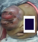
Intracranial meningiomas are usually non-cancerous tumors that develop from arachnoid cells in the meningeal envelope. However, there are rare forms called intraosseous meningiomas, which present unique challenges for diagnosis and treatment. In this report, we describe a rare case of a giant sphenotemporal meningioma in a 72-year-old male with diabetes. The patient experienced progressive exophthalmos and visual impairment over a period of five months. Radiological imaging confirmed the diagnosis, showing extensive infiltration into the infra-temporal region. Histopathological examination confirmed a plaque-type meningothelial meningioma. The patient underwent surgical management, which involved maxillofacial surgery. Intraosseous meningiomas are rare but are increasingly being recognized, accounting for about two percent of all meningiomas. The spheno-orbital region is a common site for these tumors. Histologically, there are various subtypes, with meningothelial meningioma being the most common. The differential diagnosis includes Paget’s disease and osteomas. The optimal treatment approach involves extensive surgical resection, followed by adjuvant radiotherapy for any remaining or symptomatic tumors. The prognosis depends on the extent of resection and tumor progression, underscoring the importance of regular monitoring. Early intervention is crucial to preserve visual function and achieve favorable outcomes.
Read Full Article HTML DOI: 10.29328/journal.ijceo.1001055 Cite this Article Read Full Article PDF
Keywords:
Meningioma; Exophthalmos; Orbital; Spheno-temporal
References
- Alcalá Cerra G, Moscote Salazar LR, Lozano Tangua CF, Sabogal Barrios R, García Quintana G. Primary osteolytic intra diploic meningioma: Case report. Rev Chil Neurosurgery [Internet]. 2010 [cited 2019 Dec 15]; 34(10).
- Ammirati M, Mirzai S, Samii M. Primary intraosseous meningiomas of the skull base. Acta Neurochir (Wien). 1990;107(1-2):56-60. doi: 10.1007/BF01402613. PMID: 2096610.
- Daffner RH, Yakulis R, Maroon JC. Intraosseous meningioma. Skeletal Radiol. 1998 Feb;27(2):108-11. doi: 10.1007/s002560050347. PMID: 9526778.
- Ichimura S, Hara K, Shimokawa R, Kagami H, Inaba M. A case of intraosseous microcystic meningioma without a mass lesion. Neurol Med Chir (Tokyo). 2013;53(10):699-702. doi: 10.2176/nmc.cr2012-0124. Epub 2013 Sep 24. PMID: 24064568; PMCID: PMC4508748.
- Nozaki K, Kikuta K, Takagi Y, Mineharu Y, Takahashi JA, Hashimoto N. Effect of early optic canal unroofing on the outcome of visual functions in surgery for meningiomas of the tuberculum sellae and planum sphenoidale. Neurosurgery. 2008 Apr;62(4):839-44; discussion 844-6. doi: 10.1227/01.neu.0000318169.75095.cb. PMID: 18496190.
- De Jesús O, Toledo MM. Surgical management of meningioma en plaque of the sphenoid ridge. Surg Neurol. 2001 May;55(5):265-9. doi: 10.1016/s0090-3019(01)00440-2. PMID: 11516463.
- Arregui R, Ovalle R, Castillo J. Extradural meningioma of middle ear: Reporting a case and literature review. Rev Otorrinolaringol Cir Cabeza Cuello. 2017; 77(4).
- Mariniello G, Maiuri F, de Divitiis E, Bonavolontà G, Tranfa F, Iuliano A, Strianese D. Lateral orbitotomy for removal of sphenoid wing meningiomas invading the orbit. Neurosurgery. 2010 Jun;66(6 Suppl Operative):287-92; discussion 292. doi: 10.1227/01.NEU.0000369924.87437.0B. PMID: 20489518.
- Al-Mefty O. Supraorbital-pterional approach to skull base lesions. Neurosurgery. 1987 Oct;21(4):474-7. doi: 10.1227/00006123-198710000-00006. PMID: 3683780.
- Ringel F, Cedzich C, Schramm J. Microsurgical technique and results of a series of 63 spheno-orbital meningiomas. Neurosurgery. 2007 Apr;60(4 Suppl 2):214-21; discussion 221-2. doi: 10.1227/01.NEU.0000255415.47937.1A. PMID: 17415156.
- Heufelder MJ, Sterker I, Trantakis C, Schneider JP, Meixensberger J, Hemprich A, Frerich B. Reconstructive and ophthalmologic outcomes following resection of spheno-orbital meningiomas. Ophthalmic Plast Reconstr Surg. 2009 May-Jun;25(3):223-6. doi: 10.1097/IOP.0b013e3181a1f345. PMID: 19454936.
- Van Tassel P, Lee YY, Ayala A, Carrasco CH, Klima T. Case report 680. Intraosseous meningioma of the sphenoid bone. Skeletal Radiol. 1991;20(5):383-6. doi: 10.1007/BF01267669. PMID: 1896882.
- Sandalcioglu IE, Gasser T, Mohr C, Stolke D, Wiedemayer H. Spheno-orbital meningiomas: interdisciplinary surgical approach, resectability and long-term results. J Craniomaxillofac Surg. 2005 Aug;33(4):260-6. doi: 10.1016/j.jcms.2005.01.013. PMID: 15978821.
- Brusati R, Biglioli F, Mortini P, Raffaini M, Goisis M. Reconstruction of the orbital walls in surgery of the skull base for benign neoplasms. Int J Oral Maxillofac Surg. 2000 Oct;29(5):325-30. PMID: 11071232.
Figures:
Similar Articles
-
A rare presentation of orbital dermoid: A case studySheetal V Girimallanavar*,Seema Channabasappa,Balasubrahmanyam Aluri,Divya Rose Cyriac,Aiswarya Ann Jose. A rare presentation of orbital dermoid: A case study. . 2021 doi: 10.29328/journal.ijceo.1001037; 5: 016-018
-
Exophthalmos Revealing a Spheno Temporo Orbital MeningiomaHassina S*, Krichene MA, Hazil Z, Bekkar B, Hasnaoui I, Robbana L, Bardi S, Akkanour Y, Serghini L, Abdallah EL. Exophthalmos Revealing a Spheno Temporo Orbital Meningioma. . 2024 doi: 10.29328/journal.ijceo.1001055; 8: 001-003
Recently Viewed
-
Agriculture High-Quality Development and NutritionZhongsheng Guo*. Agriculture High-Quality Development and Nutrition. Arch Food Nutr Sci. 2024: doi: 10.29328/journal.afns.1001060; 8: 038-040
-
A Low-cost High-throughput Targeted Sequencing for the Accurate Detection of Respiratory Tract PathogenChangyan Ju, Chengbosen Zhou, Zhezhi Deng, Jingwei Gao, Weizhao Jiang, Hanbing Zeng, Haiwei Huang, Yongxiang Duan, David X Deng*. A Low-cost High-throughput Targeted Sequencing for the Accurate Detection of Respiratory Tract Pathogen. Int J Clin Virol. 2024: doi: 10.29328/journal.ijcv.1001056; 8: 001-007
-
A Comparative Study of Metoprolol and Amlodipine on Mortality, Disability and Complication in Acute StrokeJayantee Kalita*,Dhiraj Kumar,Nagendra B Gutti,Sandeep K Gupta,Anadi Mishra,Vivek Singh. A Comparative Study of Metoprolol and Amlodipine on Mortality, Disability and Complication in Acute Stroke. J Neurosci Neurol Disord. 2025: doi: 10.29328/journal.jnnd.1001108; 9: 039-045
-
Development of qualitative GC MS method for simultaneous identification of PM-CCM a modified illicit drugs preparation and its modern-day application in drug-facilitated crimesBhagat Singh*,Satish R Nailkar,Chetansen A Bhadkambekar,Suneel Prajapati,Sukhminder Kaur. Development of qualitative GC MS method for simultaneous identification of PM-CCM a modified illicit drugs preparation and its modern-day application in drug-facilitated crimes. J Forensic Sci Res. 2023: doi: 10.29328/journal.jfsr.1001043; 7: 004-010
-
A Gateway to Metal Resistance: Bacterial Response to Heavy Metal Toxicity in the Biological EnvironmentLoai Aljerf*,Nuha AlMasri. A Gateway to Metal Resistance: Bacterial Response to Heavy Metal Toxicity in the Biological Environment. Ann Adv Chem. 2018: doi: 10.29328/journal.aac.1001012; 2: 032-044
Most Viewed
-
Evaluation of Biostimulants Based on Recovered Protein Hydrolysates from Animal By-products as Plant Growth EnhancersH Pérez-Aguilar*, M Lacruz-Asaro, F Arán-Ais. Evaluation of Biostimulants Based on Recovered Protein Hydrolysates from Animal By-products as Plant Growth Enhancers. J Plant Sci Phytopathol. 2023 doi: 10.29328/journal.jpsp.1001104; 7: 042-047
-
Sinonasal Myxoma Extending into the Orbit in a 4-Year Old: A Case PresentationJulian A Purrinos*, Ramzi Younis. Sinonasal Myxoma Extending into the Orbit in a 4-Year Old: A Case Presentation. Arch Case Rep. 2024 doi: 10.29328/journal.acr.1001099; 8: 075-077
-
Feasibility study of magnetic sensing for detecting single-neuron action potentialsDenis Tonini,Kai Wu,Renata Saha,Jian-Ping Wang*. Feasibility study of magnetic sensing for detecting single-neuron action potentials. Ann Biomed Sci Eng. 2022 doi: 10.29328/journal.abse.1001018; 6: 019-029
-
Pediatric Dysgerminoma: Unveiling a Rare Ovarian TumorFaten Limaiem*, Khalil Saffar, Ahmed Halouani. Pediatric Dysgerminoma: Unveiling a Rare Ovarian Tumor. Arch Case Rep. 2024 doi: 10.29328/journal.acr.1001087; 8: 010-013
-
Physical activity can change the physiological and psychological circumstances during COVID-19 pandemic: A narrative reviewKhashayar Maroufi*. Physical activity can change the physiological and psychological circumstances during COVID-19 pandemic: A narrative review. J Sports Med Ther. 2021 doi: 10.29328/journal.jsmt.1001051; 6: 001-007

HSPI: We're glad you're here. Please click "create a new Query" if you are a new visitor to our website and need further information from us.
If you are already a member of our network and need to keep track of any developments regarding a question you have already submitted, click "take me to my Query."


