Clinical Image
Chronic recurrent bilateral granulomatous iridocyclitis in an 18-year-old woman

Marie-Hélène Errera1*, Cameron F Parsa2, Jonathan Benesty2, Neila Sedira2, Raphaël Thouvenin2, Emmanuel Héron2 and José-Alain Sahel2
1MD, Centre Quinze-Vingts National Ophthalmology Hospital, 28, Rue de Charenton, 75012 Paris, France2MD, Quinze-Vingts National Eye Hospital, UPMC-Sorbonne University, Paris, France
*Address for Correspondence: Marie-Hélène Errera, MD, Centre Quinze-Vingts National Ophthalmology Hospital, 28, Rue de Charenton, 75012 Paris, France, Tel: +33 1 40 02 14 15; Email: [email protected]
Dates: Submitted: 30 July 2018; Approved: 30 July 2018; Published: 31 July 2018
How to cite this article: Errera MH, Parsa CF, Benesty J, Sedira N, Thouvenin R, et al. Chronic recurrent bilateral granulomatous iridocyclitis in an 18-year-old woman. Int J Clin Exp Ophthalmol. 2018; 2: 021-021. DOI: 10.29328/journal.ijceo.1001016
Copyright Licence: © 2018 Errera MH, et al. This is an open access article distributed under the Creative Commons Attribution License, which permits unrestricted use, distribution, and reproduction in any medium, provided the original work is properly cited.
Clinical Image
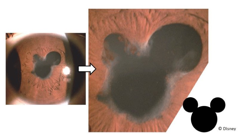
A case of recurrent tuberculosis-related uveitis in a young female patient of North African origin.
Right eye: Slit-lamp photograph of broad-based posterior synechiae (formed by macrophage-laden iris tissue when brushing against and then adhering to, the anterior lens capsule) rendering the pupils irregular, clover- or, here, “Mickey Mouse”©-shaped. Cycloplegic agents can increase the pupillary circumference, displacing iris tissue further away from the crystalline lens to help reduce adhesions from forming that beget lens opacities as well as block aqueous humor flow.
Figure 1: A case of recurrent tuberculosis-related uveitis in a young female patient of North African origin. Right eye: Slit-lamp photograph of broad-based posterior synechiae (formed by macrophage-laden iris tissue when brushing against and then adhering to, the anterior lens capsule) rendering the pupils irregular, clover- or, here, “Mickey Mouse”©-shaped. Cycloplegic agents can increase the pupillary circumference, displacing iris tissue further away from the crystalline lens to help reduce adhesions from forming that beget lens opacities as well as block aqueous humor flow.

