Figure 1
Treatment of Retinal Pigment Epithelial Detachment
Ayse Gul Kocak Altintas* and Cagri Ilhan
Published: 23 March, 2018 | Volume 2 - Issue 1 | Pages: 008-014

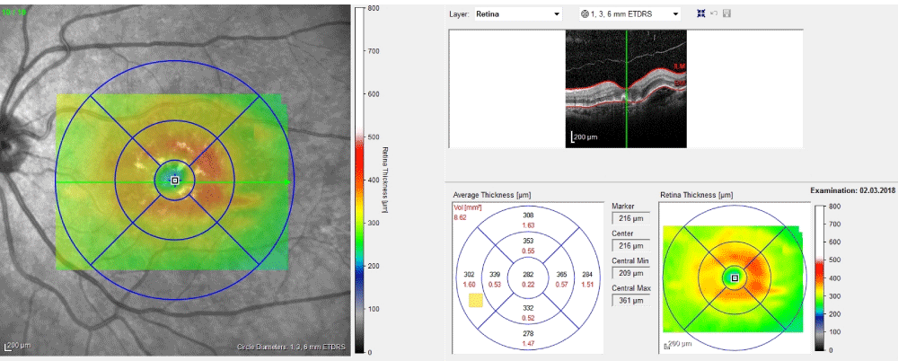
Figure 1:
Macular thickness map of drusenoid pigment epithelial detachment.
Read Full Article HTML DOI: 10.29328/journal.ijceo.1001013 Cite this Article Read Full Article PDF
More Images
Similar Articles
-
Intravitreal Ranibizumab/ Lucentis (IVTL) injections in Glaucoma patients-Intraocular Pressure (IOP) elevation and the use of Anterior Chamber Paracentesis (ACP)EA Ansari*. Intravitreal Ranibizumab/ Lucentis (IVTL) injections in Glaucoma patients-Intraocular Pressure (IOP) elevation and the use of Anterior Chamber Paracentesis (ACP) . . 2017 doi: 10.29328/journal.hceo.1001005; 1: 033-041
-
Intravitreal ranibizumab in the management of acute central serous ChorioretinopathyIbrahim Nawaiseh,Ahmad Halawa*,Dina Alardah. Intravitreal ranibizumab in the management of acute central serous Chorioretinopathy . . 2017 doi: 10.29328/journal.hceo.1001007; 1: 049-054
-
Treatment of Retinal Pigment Epithelial DetachmentAyse Gul Kocak Altintas*,Cagri Ilhan. Treatment of Retinal Pigment Epithelial Detachment . . 2018 doi: 10.29328/journal.ijceo.1001013; 2: 008-014
Recently Viewed
-
Late discover of a traumatic cardiac injury: Case reportBenlafqih C,Bouhdadi H*,Bakkali A,Rhissassi J,Sayah R,Laaroussi M. Late discover of a traumatic cardiac injury: Case report. J Cardiol Cardiovasc Med. 2019: doi: 10.29328/journal.jccm.1001048; 4: 100-102
-
A two-phase sonographic study among women with infertility who first had normal sonographic findingsKalu Ochie*,Abraham John C. A two-phase sonographic study among women with infertility who first had normal sonographic findings. Clin J Obstet Gynecol. 2022: doi: 10.29328/journal.cjog.1001117; 5: 101-103
-
Sinonasal Myxoma Extending into the Orbit in a 4-Year Old: A Case PresentationJulian A Purrinos*, Ramzi Younis. Sinonasal Myxoma Extending into the Orbit in a 4-Year Old: A Case Presentation. Arch Case Rep. 2024: doi: 10.29328/journal.acr.1001099; 8: 075-077
-
Analysis of Psychological and Physiological Responses to Snoezelen Multisensory StimulationLucia Ludvigh Cintulova,Jerzy Rottermund,Zuzana Budayova. Analysis of Psychological and Physiological Responses to Snoezelen Multisensory Stimulation. J Neurosci Neurol Disord. 2024: doi: 10.29328/journal.jnnd.1001103; 8: 115-125
-
Feature Processing Methods: Recent Advances and Future TrendsShiying Bai,Lufeng Bai*. Feature Processing Methods: Recent Advances and Future Trends. J Clin Med Exp Images. 2025: doi: 10.29328/journal.jcmei.1001035; 9: 010-014
Most Viewed
-
Evaluation of Biostimulants Based on Recovered Protein Hydrolysates from Animal By-products as Plant Growth EnhancersH Pérez-Aguilar*, M Lacruz-Asaro, F Arán-Ais. Evaluation of Biostimulants Based on Recovered Protein Hydrolysates from Animal By-products as Plant Growth Enhancers. J Plant Sci Phytopathol. 2023 doi: 10.29328/journal.jpsp.1001104; 7: 042-047
-
Sinonasal Myxoma Extending into the Orbit in a 4-Year Old: A Case PresentationJulian A Purrinos*, Ramzi Younis. Sinonasal Myxoma Extending into the Orbit in a 4-Year Old: A Case Presentation. Arch Case Rep. 2024 doi: 10.29328/journal.acr.1001099; 8: 075-077
-
Feasibility study of magnetic sensing for detecting single-neuron action potentialsDenis Tonini,Kai Wu,Renata Saha,Jian-Ping Wang*. Feasibility study of magnetic sensing for detecting single-neuron action potentials. Ann Biomed Sci Eng. 2022 doi: 10.29328/journal.abse.1001018; 6: 019-029
-
Pediatric Dysgerminoma: Unveiling a Rare Ovarian TumorFaten Limaiem*, Khalil Saffar, Ahmed Halouani. Pediatric Dysgerminoma: Unveiling a Rare Ovarian Tumor. Arch Case Rep. 2024 doi: 10.29328/journal.acr.1001087; 8: 010-013
-
Physical activity can change the physiological and psychological circumstances during COVID-19 pandemic: A narrative reviewKhashayar Maroufi*. Physical activity can change the physiological and psychological circumstances during COVID-19 pandemic: A narrative review. J Sports Med Ther. 2021 doi: 10.29328/journal.jsmt.1001051; 6: 001-007

HSPI: We're glad you're here. Please click "create a new Query" if you are a new visitor to our website and need further information from us.
If you are already a member of our network and need to keep track of any developments regarding a question you have already submitted, click "take me to my Query."


