Figure 1
Chronic recurrent bilateral granulomatous iridocyclitis in an 18-year-old woman
Marie-Hélène Errera*, Cameron F Parsa, Jonathan Benesty, Neila Sedira, Raphaël Thouvenin, Emmanuel Héron and José-Alain Sahel
Published: 31 July, 2018 | Volume 2 - Issue 2 | Pages: 021-021
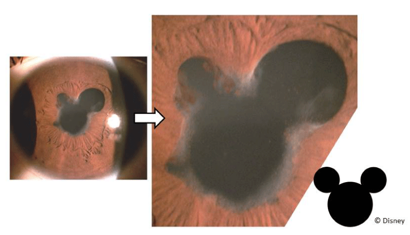
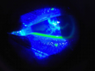
Figure 1:
A case of recurrent tuberculosis-related uveitis in a young female patient of North African origin. Right eye: Slit-lamp photograph of broad-based posterior synechiae (formed by macrophage-laden iris tissue when brushing against and then adhering to, the anterior lens capsule) rendering the pupils irregular, clover- or, here, “Mickey Mouse”©-shaped. Cycloplegic agents can increase the pupillary circumference, displacing iris tissue further away from the crystalline lens to help reduce adhesions from forming that beget lens opacities as well as block aqueous humor flow.
Read Full Article HTML DOI: 10.29328/journal.ijceo.1001016 Cite this Article Read Full Article PDF
More Images
Similar Articles
-
A Comparative Study of Anatomic and Functional Outcomes of Two Surgical Techniques of Cataract at LomeAyena KD*,Santos KAM,Vonor K,Amedome KM,Wodome A,Strauss G,Nagbe YE,Koffi-Ametooyona A, Balo K. A Comparative Study of Anatomic and Functional Outcomes of Two Surgical Techniques of Cataract at Lome . . 2017 doi: 10.29328/journal.hceo.1001001; 1: 001-008
-
Theory and Experiments. (+) Add Reading Glasses to Prevent MyopiaPeter R Greene*,Brown OS. Theory and Experiments. (+) Add Reading Glasses to Prevent Myopia . . 2017 doi: 10.29328/journal.hceo.1001002; 1: 009-022
-
Efficacy of early Hyperbaric Oxygen Therapy in Central Retinal Artery OcclusionAyse Gul Kocak Altintas*,Mehmet Citirik. Efficacy of early Hyperbaric Oxygen Therapy in Central Retinal Artery Occlusion . . 2017 doi: 10.29328/journal.hceo.1001003; 1: 023-028
-
Mitomycin-C Use and Complications in OphthalmologyTongabay Cumurcu*. Mitomycin-C Use and Complications in Ophthalmology. . 2017 doi: 10.29328/journal.hceo.1001004; 1: 029-032
-
Intravitreal Ranibizumab/ Lucentis (IVTL) injections in Glaucoma patients-Intraocular Pressure (IOP) elevation and the use of Anterior Chamber Paracentesis (ACP)EA Ansari*. Intravitreal Ranibizumab/ Lucentis (IVTL) injections in Glaucoma patients-Intraocular Pressure (IOP) elevation and the use of Anterior Chamber Paracentesis (ACP) . . 2017 doi: 10.29328/journal.hceo.1001005; 1: 033-041
-
Detection of Ganglion Cell Loss in Preperimetric Glaucoma by Fourier-Domain Optical Coherence TomographySuneeta Dubey*, Baswati Prasanth,Lokesh Chauhan, Saptarshi Mukherjee. Detection of Ganglion Cell Loss in Preperimetric Glaucoma by Fourier-Domain Optical Coherence Tomography. . 2017 doi: 10.29328/journal.hceo.1001006; 1: 042-048
-
Intravitreal ranibizumab in the management of acute central serous ChorioretinopathyIbrahim Nawaiseh,Ahmad Halawa*,Dina Alardah. Intravitreal ranibizumab in the management of acute central serous Chorioretinopathy . . 2017 doi: 10.29328/journal.hceo.1001007; 1: 049-054
-
The Role of Omega-3 Essential Fatty Acids in Dry Eye DiseaseWilliam J Faulkner*. The Role of Omega-3 Essential Fatty Acids in Dry Eye Disease . . 2017 doi: 10.29328/journal.ijceo.1001008; 1: 055-059
-
Neuro-ophthalmological emergency disorders: A general viewBurak Turgut*,Feyza Çaliş Karanfil, Fatoş Altun Turgut. Neuro-ophthalmological emergency disorders: A general view . . 2017 doi: 10.29328/journal.ijceo.1001009; 1: 060-066
-
Prospective Clinical Study to Find out Epidemiology of Xerophthalmia in Children in a Tertiary Care Centre in IndiaDeepak Mishra*,Megha Gulati,Prashant Bhushan,Nilesh Mohan,Bibhuti Sinha P. Prospective Clinical Study to Find out Epidemiology of Xerophthalmia in Children in a Tertiary Care Centre in India . . 2017 doi: 10.29328/journal.ijceo.1001010; 1: 066-070
Recently Viewed
-
GELS as Pharmaceutical Form in Hospital Galenic Practice: Chemico-physical and Pharmaceutical AspectsLuisetto M*,Edbey Kaled,Mashori GR,Ferraiuolo A,Fiazza C,Cabianca L,Latyschev OY. GELS as Pharmaceutical Form in Hospital Galenic Practice: Chemico-physical and Pharmaceutical Aspects. Arch Surg Clin Res. 2025: doi: 10.29328/journal.ascr.1001084; 9: 001-007
-
Septic Shock on Bartholinitis: Case Report and Modern Surgical ApproachesOumaima Fakir*,Hanaa Lazhar,Aziz Slaoui,Amina Lakhdar,Aziz Baydada. Septic Shock on Bartholinitis: Case Report and Modern Surgical Approaches. Clin J Obstet Gynecol. 2025: doi: 10.29328/journal.cjog.1001183; 8: 015-018
-
A Comparative Study of Metoprolol and Amlodipine on Mortality, Disability and Complication in Acute StrokeJayantee Kalita*,Dhiraj Kumar,Nagendra B Gutti,Sandeep K Gupta,Anadi Mishra,Vivek Singh. A Comparative Study of Metoprolol and Amlodipine on Mortality, Disability and Complication in Acute Stroke. J Neurosci Neurol Disord. 2025: doi: 10.29328/journal.jnnd.1001108; 9: 039-045
-
An Observational Study on Clinical Outcome and Predictors of Traumatic Cervical Injury at a Tertiary Care FacilitySurendra Saini,Ankit Prajapati,Ina Bahl*,Deepika Gehlot,Namita Prajapati. An Observational Study on Clinical Outcome and Predictors of Traumatic Cervical Injury at a Tertiary Care Facility. J Neurosci Neurol Disord. 2025: doi: 10.29328/journal.jnnd.1001107; 9: 028-038
-
Imaging modalities delivery of RNAi therapeutics in cancer therapy and clinical applicationsLoutfy H Madkour*. Imaging modalities delivery of RNAi therapeutics in cancer therapy and clinical applications. J Radiol Oncol. 2021: doi: 10.29328/journal.jro.1001035; 5: 005-034
Most Viewed
-
Evaluation of Biostimulants Based on Recovered Protein Hydrolysates from Animal By-products as Plant Growth EnhancersH Pérez-Aguilar*, M Lacruz-Asaro, F Arán-Ais. Evaluation of Biostimulants Based on Recovered Protein Hydrolysates from Animal By-products as Plant Growth Enhancers. J Plant Sci Phytopathol. 2023 doi: 10.29328/journal.jpsp.1001104; 7: 042-047
-
Sinonasal Myxoma Extending into the Orbit in a 4-Year Old: A Case PresentationJulian A Purrinos*, Ramzi Younis. Sinonasal Myxoma Extending into the Orbit in a 4-Year Old: A Case Presentation. Arch Case Rep. 2024 doi: 10.29328/journal.acr.1001099; 8: 075-077
-
Feasibility study of magnetic sensing for detecting single-neuron action potentialsDenis Tonini,Kai Wu,Renata Saha,Jian-Ping Wang*. Feasibility study of magnetic sensing for detecting single-neuron action potentials. Ann Biomed Sci Eng. 2022 doi: 10.29328/journal.abse.1001018; 6: 019-029
-
Pediatric Dysgerminoma: Unveiling a Rare Ovarian TumorFaten Limaiem*, Khalil Saffar, Ahmed Halouani. Pediatric Dysgerminoma: Unveiling a Rare Ovarian Tumor. Arch Case Rep. 2024 doi: 10.29328/journal.acr.1001087; 8: 010-013
-
Physical activity can change the physiological and psychological circumstances during COVID-19 pandemic: A narrative reviewKhashayar Maroufi*. Physical activity can change the physiological and psychological circumstances during COVID-19 pandemic: A narrative review. J Sports Med Ther. 2021 doi: 10.29328/journal.jsmt.1001051; 6: 001-007

HSPI: We're glad you're here. Please click "create a new Query" if you are a new visitor to our website and need further information from us.
If you are already a member of our network and need to keep track of any developments regarding a question you have already submitted, click "take me to my Query."


