Figure 5
Unilateral retinitis pigmentosa: Case report and review of the literature
Denise Goodwin*, Amanda M Olsen and Karl Citek
Published: 22 August, 2019 | Volume 3 - Issue 2 | Pages: 013-19
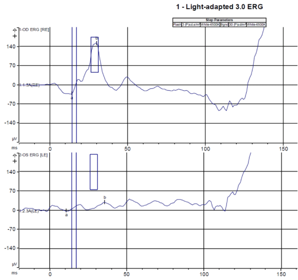
Figure 5:
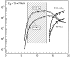
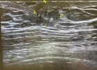
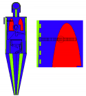
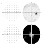
Full field electroretinography showed normal findings in the right eye and significantly reduced function in the left eye with the light-adapted 3.0 electroretinography (a), light-adapted 3.0 flicker electroretinography (b), dark-adapted 0.01 electroretinography (c), dark-adapted 3.0 electroretinography (d), and dark-adapted 10.0 electroretinography.
Read Full Article HTML DOI: 10.29328/journal.ijceo.1001021 Cite this Article Read Full Article PDF
More Images
Similar Articles
-
Detection of Ganglion Cell Loss in Preperimetric Glaucoma by Fourier-Domain Optical Coherence TomographySuneeta Dubey*, Baswati Prasanth,Lokesh Chauhan, Saptarshi Mukherjee. Detection of Ganglion Cell Loss in Preperimetric Glaucoma by Fourier-Domain Optical Coherence Tomography. . 2017 doi: 10.29328/journal.hceo.1001006; 1: 042-048
-
Intravitreal ranibizumab in the management of acute central serous ChorioretinopathyIbrahim Nawaiseh,Ahmad Halawa*,Dina Alardah. Intravitreal ranibizumab in the management of acute central serous Chorioretinopathy . . 2017 doi: 10.29328/journal.hceo.1001007; 1: 049-054
-
The management of Irvine-Gass Syndrome in a patient using Inhaler SteroidAyse Gul Kocak Altintas*,Cagri Ilhan,Mehmet Citirik. The management of Irvine-Gass Syndrome in a patient using Inhaler Steroid . . 2018 doi: 10.29328/journal.ijceo.1001011; 2: 001-005
-
Treatment of Retinal Pigment Epithelial DetachmentAyse Gul Kocak Altintas*,Cagri Ilhan. Treatment of Retinal Pigment Epithelial Detachment . . 2018 doi: 10.29328/journal.ijceo.1001013; 2: 008-014
-
Low vision due to persistent Cloquet’s canalAyse Gul Kocak Altintas*. Low vision due to persistent Cloquet’s canal . . 2018 doi: 10.29328/journal.ijceo.1001014; 2: 015-017
-
Wound architectural analysis of 1.8mm microincision cataract surgery using spectral domain OCTBenedito Antônio de Sousa, Anderson Teixeira*,Camila Salaroli,Nonato Souza,Lucy Gomes. Wound architectural analysis of 1.8mm microincision cataract surgery using spectral domain OCT. . 2019 doi: 10.29328/journal.ijceo.1001020; 3: 008-012
-
Unilateral retinitis pigmentosa: Case report and review of the literatureDenise Goodwin*,Amanda M Olsen,Karl Citek. Unilateral retinitis pigmentosa: Case report and review of the literature. . 2019 doi: 10.29328/journal.ijceo.1001021; 3: 013-19
-
Evaluation of the efficacy of transcorneal electric stimulation therapy in retinitis pigmentosa patients with electrophysiological and structural testsMualla Sahin Hamurcu*,Sema Akkan Aydogmuş,M. Sinan SARICAOĞLU. Evaluation of the efficacy of transcorneal electric stimulation therapy in retinitis pigmentosa patients with electrophysiological and structural tests. . 2020 doi: 10.29328/journal.ijceo.1001030; 4: 031-037
-
Can we predict Alzheimer’s Disease through the eye lens?Sandeep CS*,Vijayakumar N, Sukesh Kumar A. Can we predict Alzheimer’s Disease through the eye lens?. . 2020 doi: 10.29328/journal.ijceo.1001031; 4: 038-040
-
Clinical evaluation of the eye-to-visual-pathway integrity of glaucomatous neurodegeneration using 1.5T MR imaging: The integrity of glaucomatous neurodegenerationKaya N Engin*,Ulviye Yiğit#,Sibel Töreyen Bayramoğlu#,Nurten Turan Güner#,Onur Özyurt#,Kutlay Tufan#,Ahmet Ağaçhan#,Penbe Çağatay#. Clinical evaluation of the eye-to-visual-pathway integrity of glaucomatous neurodegeneration using 1.5T MR imaging: The integrity of glaucomatous neurodegeneration. . 2022 doi: 10.29328/journal.ijceo.1001042; 6: 001-006
Recently Viewed
-
Potential Use of Essential Oils and Their Individual Components in Cosmeceuticals: A ReviewHamdy A Shaaban*. Potential Use of Essential Oils and Their Individual Components in Cosmeceuticals: A Review. Ann Biomed Sci Eng. 2023: doi: 10.29328/journal.abse.1001023; 7: 031-037
-
Studies on some spices and herbs: Chemical composition, health benefits and functional propertiesHamdy A Shaaban*. Studies on some spices and herbs: Chemical composition, health benefits and functional properties. Ann Biomed Sci Eng. 2023: doi: 10.29328/journal.abse.1001019; 7: 001-011
-
About Efficiency of High-order Harmonic Generation in Attosecond PhysicsAng-Yang Yu*. About Efficiency of High-order Harmonic Generation in Attosecond Physics. Int J Clin Virol. 2024: doi: 10.29328/journal.ijcv.1001061; 8: 045-047
-
Success, Survival and Prognostic Factors in Implant Prosthesis: Experimental StudyEpifania Ettore*, Pietrantonio Maria, Christian Nunziata, Ausiello Pietro. Success, Survival and Prognostic Factors in Implant Prosthesis: Experimental Study. J Oral Health Craniofac Sci. 2023: doi: 10.29328/journal.johcs.1001045; 8: 024-028
-
Agriculture High-Quality Development and NutritionZhongsheng Guo*. Agriculture High-Quality Development and Nutrition. Arch Food Nutr Sci. 2024: doi: 10.29328/journal.afns.1001060; 8: 038-040
Most Viewed
-
Evaluation of Biostimulants Based on Recovered Protein Hydrolysates from Animal By-products as Plant Growth EnhancersH Pérez-Aguilar*, M Lacruz-Asaro, F Arán-Ais. Evaluation of Biostimulants Based on Recovered Protein Hydrolysates from Animal By-products as Plant Growth Enhancers. J Plant Sci Phytopathol. 2023 doi: 10.29328/journal.jpsp.1001104; 7: 042-047
-
Sinonasal Myxoma Extending into the Orbit in a 4-Year Old: A Case PresentationJulian A Purrinos*, Ramzi Younis. Sinonasal Myxoma Extending into the Orbit in a 4-Year Old: A Case Presentation. Arch Case Rep. 2024 doi: 10.29328/journal.acr.1001099; 8: 075-077
-
Feasibility study of magnetic sensing for detecting single-neuron action potentialsDenis Tonini,Kai Wu,Renata Saha,Jian-Ping Wang*. Feasibility study of magnetic sensing for detecting single-neuron action potentials. Ann Biomed Sci Eng. 2022 doi: 10.29328/journal.abse.1001018; 6: 019-029
-
Pediatric Dysgerminoma: Unveiling a Rare Ovarian TumorFaten Limaiem*, Khalil Saffar, Ahmed Halouani. Pediatric Dysgerminoma: Unveiling a Rare Ovarian Tumor. Arch Case Rep. 2024 doi: 10.29328/journal.acr.1001087; 8: 010-013
-
Physical activity can change the physiological and psychological circumstances during COVID-19 pandemic: A narrative reviewKhashayar Maroufi*. Physical activity can change the physiological and psychological circumstances during COVID-19 pandemic: A narrative review. J Sports Med Ther. 2021 doi: 10.29328/journal.jsmt.1001051; 6: 001-007

HSPI: We're glad you're here. Please click "create a new Query" if you are a new visitor to our website and need further information from us.
If you are already a member of our network and need to keep track of any developments regarding a question you have already submitted, click "take me to my Query."


